
A Modern Guide to the Restriction Analysis Protocol
Team Meloq
Author
As clinicians, our goal is to move beyond subjective observation to objective measurement when evaluating a patient's progress. A restriction analysis protocol offers a structured, data-driven method for this purpose.
It is a systematic way to measure real-world physical limitations. Instead of relying on visual estimation, this protocol uses reliable tools to gather quantifiable data on a patient's range of motion, muscle strength, and balance. This evidence-based approach can lead to more precise diagnoses and personalized treatment plans, moving clinical practice towards a more scientific foundation.
Translating Scientific Precision into Clinical Practice
The term 'restriction analysis' may be familiar to those with a background in genetics. The parallel is intentional, as it highlights a shared philosophy: the shift from subjective assessment to objective, repeatable measurement.
Just as geneticists developed reliable methods to map DNA, clinicians require standardized protocols to map and understand human movement.
This modern protocol is about bringing a high level of precision into daily clinical work. It’s a systematic approach designed to answer critical questions with concrete numbers: How limited is a joint's mobility? How much has strength improved since the last session? Is there a significant asymmetry between limbs that could increase the risk of re-injury?
From Genetics to Biomechanics
The concept of precise, protocol-driven analysis has a rich history in science. In the 1950s, Salvador Luria's work on 'host-controlled restriction' laid the groundwork for understanding how bacteria defend against viruses. By 1978, this foundational science enabled Genentech to use restriction enzymes for producing human insulin in bacteria, a breakthrough that significantly reduced treatment costs for individuals with diabetes.
This history illustrates the profound real-world impact of precise, standardized protocols. We apply the same principle in physiotherapy and rehabilitation: replacing subjective descriptions with data-driven protocols. This shift is powered by a commitment to evidence-based practice, providing a clear, objective picture of a patient's functional status.
The Core Principles of a Modern Protocol
A well-designed restriction analysis protocol is built on key pillars that ensure the data collected is both reliable and clinically useful.
- Objectivity: Measurements are taken with calibrated digital tools, which reduces the subjective judgment of the clinician.
- Reproducibility: The protocol is standardized. This means the same test performed by different therapists or on different days should yield comparable results, assuming no clinical change has occurred.
- Quantification: Instead of describing movement as "limited" or "weak," the protocol provides specific numbers (e.g., degrees of motion or Newtons of force).
By adhering to these principles, assessments can be transformed from simple observation to objective measurement. This forms the foundation for creating more effective and personalized treatment plans that can lead to better patient outcomes.
Modern advancements like mobile app development for healthcare are making it easier to implement this precision in the clinic. These digital tools are valuable for capturing, analyzing, and sharing data efficiently.
Building Your Toolkit for Objective Assessment
To execute a reliable restriction analysis protocol, the quality of your tools is paramount. The goal is to transition from subjective observation to collecting objective data. This requires instruments that deliver accurate and consistent measurements.
This is not about acquiring the most expensive equipment, but rather about building a foundation of trust in the data collected. The right toolkit helps ensure that the data gathered today is directly comparable to what will be found next week or next month. This consistency provides a clear, unbiased view of a client’s progress.
Core Instruments for Quantified Analysis
While a specific clinical focus will shape the toolkit, several core instruments are essential for any comprehensive assessment. These devices are designed to capture the key pillars of movement health—motion, strength, and balance—with digital precision.
-
Digital Goniometers: These have become a modern standard for measuring Range of Motion (ROM). They help eliminate the guesswork and parallax error that can affect traditional plastic goniometers, providing degree-specific accuracy that is valuable for tracking subtle but clinically significant changes in joint mobility.
-
Handheld Dynamometers: For quantifiable Manual Muscle Testing (MMT), a handheld dynamometer is a key tool. Instead of a subjective 0-5 scale, these devices provide precise force output in units such as Newtons. This allows for accurate strength measurement, identification of asymmetries, and the setting of objective, data-driven rehabilitation goals.
-
Force Plates: For assessing balance and lower-limb asymmetries, force plates offer a depth of insight that visual observation cannot match. They provide objective data on weight distribution, stability, and power output during functional movements.
These tools are not just for collecting data; they are for collecting reliable data. The aim is to minimize both inter-rater and intra-rater variability—inconsistencies that can undermine clinical findings. Digital instruments are engineered to address this challenge.
Precision Inspired by Science
The drive for precision in clinical practice mirrors advancements in other scientific fields. The formal classification of restriction-modification (R-M) systems in genetics, for instance, created a protocol framework that accelerated genomic research. Type II restriction enzymes can cleave DNA at specific sites with remarkable accuracy. This level of precision was transformative for various industries.
In a clinical setting, that same principle of precision is now accessible through modern tools. A handheld dynamometer, for example, can quantify peak force with high reliability. This level of accuracy allows for the replacement of subjective muscle testing with standardized, objective metrics, which can lead to better-documented and potentially more efficient patient recovery.
Selecting and Maintaining Your Equipment
Choosing the right equipment is a critical first step. Look for devices with validated accuracy and high reliability, preferably supported by clinical studies. Practical factors such as portability, ease of use, and software integration for data capture and analysis are also important considerations.
A tool is only as good as its calibration. Regularly check your devices against manufacturer guidelines to ensure they remain accurate. A simple, consistent check-in can prevent data drift and maintain the integrity of your long-term patient records.
You can explore a detailed guide on what to look for in modern force measurement equipment.
By investing in quality tools and establishing a routine for their upkeep, you build a system that produces data you can trust. This empowers you to make more confident clinical decisions, demonstrate progress to patients with clear evidence, and elevate the standard of care in your practice.
Putting the Movement Analysis Protocol into Action
With a toolkit ready, it's time to move from theory to practice. A restriction analysis protocol comes alive during its structured, real-world application. Successful execution depends on standardization—every detail, from patient positioning to verbal cues, must be consistent to generate reliable and clinically meaningful data.
This process is a systematic investigation into the three core pillars of movement health: range of motion, muscle strength, and functional symmetry. By taking a methodical approach to each, you can develop a clear, objective picture of a patient's status.
This process flow shows how the essential components come together for a comprehensive analysis.
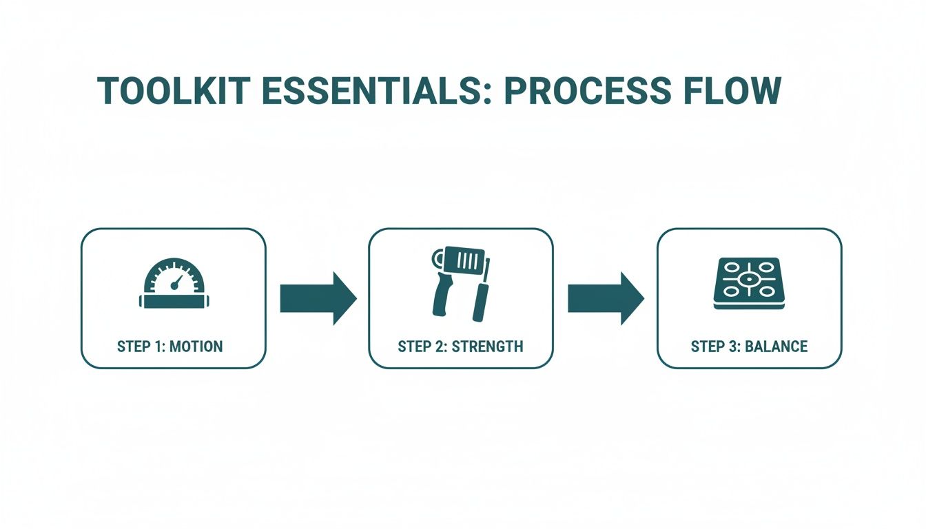
As shown, the assessment can flow from evaluating motion with a goniometer, to quantifying strength with a dynamometer, and analyzing balance and symmetry using a force plate.
Here is a quick overview of the key components involved in a standardized protocol.
Key Components Of A Standardized Restriction Analysis Protocol
| Assessment Pillar | Primary Metric | Recommended Tool | Key Standardization Tip |
|---|---|---|---|
| Motion | Degrees of joint movement | Digital Goniometer | Stabilize adjacent joints to prevent compensatory movements. |
| Strength | Peak isometric force (Newtons) | Handheld Dynamometer | Use consistent joint angles and verbal cues for every test. |
| Asymmetry | Limb Symmetry Index (LSI) | Dynamometer/Force Plate | Test the unaffected limb first to establish a reliable baseline. |
This table serves as a quick-reference guide. Let's explore how to execute each pillar with precision.
Analyzing Range Of Motion With Precision
The first pillar is quantifying joint mobility. Accurately measuring range of motion (ROM) is fundamental, as even small limitations can have significant functional consequences. The goal is to isolate the specific joint movement while preventing compensatory movements that could affect the measurement.
Proper patient positioning is non-negotiable. For example, when assessing glenohumeral internal rotation, the patient should be supine with their scapula stabilized against the table. This step helps prevent thoracic rotation or scapular tilting from artificially inflating the measurement.
Once positioned, use a digital goniometer to align with the correct anatomical landmarks before moving the limb passively to its end range. It is important to apply consistent pressure to find the true end-feel without causing unnecessary discomfort. Documenting the exact degrees provides an objective baseline that subjective terms like "mildly restricted" cannot match.
Don't underestimate the power of verbal cues. Simple, clear instructions like "relax completely and let me move your arm" help ensure the patient isn't actively assisting or resisting. This is essential for a true passive ROM measurement. Consistency in cues is as important as consistency in positioning.
Quantifying Muscle Strength Objectively
Next, let's assign a number to muscle strength. Traditional manual muscle testing (MMT) and its subjective grading scale have long been clinical staples, but a modern restriction analysis protocol demands more precision. A handheld dynamometer is indispensable for this.
Consider a patient recovering from an ACL reconstruction. A critical metric for a safe return to sport is the Limb Symmetry Index (LSI), which compares the quadriceps strength of the involved leg to the uninvolved one. To measure this, you would perform an isometric "make" test.
The procedure must be strictly standardized:
- Positioning: Seat the patient securely with their knee bent to a specific, repeatable angle (e.g., 90 degrees).
- Stabilization: Ensure their pelvis is stable and they are not using their hands for bracing, which can alter force output.
- Application: Place the dynamometer on their anterior tibia and instruct them to "kick out as hard as you can for five seconds."
By recording the peak force in Newtons from both limbs, you can calculate the LSI. Some research suggests that an LSI below 90% may be associated with a higher risk of re-injury, providing a data-driven benchmark to guide rehabilitation (1). For a deeper dive, you can read more about the analysis of movement in our other guides.
Identifying Asymmetries In Function
The final pillar is assessing asymmetries, which can expose underlying neuromuscular deficits or compensatory strategies that may increase injury risk. While a strength LSI is one way to examine asymmetry, force plates can provide a richer, more dynamic picture.
For instance, when assessing a basketball player's jump landing mechanics, a drop jump onto force plates can reveal asymmetries in ground reaction forces, loading rates, and time to stabilization between their left and right legs.
This objective data might show that the athlete consistently lands with a significantly greater proportion of their force on one leg—a risky pattern that visual observation could miss. This provides a specific, actionable target for intervention, such as focusing on single-leg neuromuscular control or targeted strengthening for the weaker side. This level of detail elevates the assessment from a general screen to a precise diagnostic tool, allowing for more targeted interventions.
Making the Numbers Mean Something: From Raw Data to Actionable Insights
Collecting accurate numbers with a goniometer or dynamometer is a crucial first step, but it's only part of the process. The clinical skill lies in turning raw measurements into a coherent narrative that guides treatment. This involves connecting a specific degree of motion to a patient's real-world ability to perform daily tasks, or linking a force deficit to their risk of future injury.

This process is about more than just data entry. It's about building a story of progress, pinpointing meaningful changes, and using objective data to educate patients and encourage their participation. A well-interpreted dataset elevates the clinician from a service provider to a trusted advisor backed by solid evidence.
Nailing Down the Clinical Baseline
Before tracking progress, a solid starting point is needed. The initial assessment is the opportunity to establish a comprehensive baseline for every key metric. This is not just a snapshot; it's the foundation against which all future measurements will be judged.
For a post-operative ACL patient on day one, a baseline assessment might capture:
- Knee flexion and extension ROM: Measured precisely to the degree.
- Quadriceps and hamstring peak force: Quantified in Newtons.
- Single-leg balance: Timed or measured with sway data from a force plate.
This baseline becomes an objective anchor. When the patient returns in two weeks, you can compare their new peak force of 220N to their initial 185N, providing clear proof of change and moving beyond subjective impressions.
Giving Your Findings Context with Normative Data
A patient's data becomes more powerful when placed in a broader context. Is a 15% limb symmetry deficit in hamstring strength significant for a professional soccer player? How does a 55-year-old’s grip strength compare to the average for their age and gender? This is where normative data and clinical benchmarks are valuable.
By comparing your patient’s numbers to established norms, you can classify the severity of a deficit and set realistic, evidence-based goals for their recovery. It adds a layer of scientific rigor to your clinical reasoning.
For instance, research suggests that athletes with a hamstring-to-quadriceps strength ratio below a certain threshold may be at a higher risk for hamstring strains (2). If your assessment shows a ratio below this benchmark, you now have a compelling, evidence-based reason to focus on posterior chain strengthening. The recommendation transforms from a general suggestion into a targeted, risk-reduction strategy.
Spotting a Clinically Significant Change
Not every fluctuation in measurements is meaningful. A patient's ROM might vary by a degree or two between sessions due to normal daily variation. The skill is in identifying the minimal clinically important difference (MCID)—the smallest change in an outcome that a patient would perceive as beneficial.
Knowing the MCID for your tests helps you avoid overreacting to minor data noise. It allows you to say with confidence, "Your shoulder external rotation has improved by 12 degrees, which is a significant change that should make reaching into a high cabinet much easier."
This focus on statistical rigor is not unique to this field. The out-of-specification (OOS) investigation protocols in the biopharmaceutical industry, which were influenced by early quality control methods, recognize that random "out-of-spec" results can occur due to normal statistical variation. Their strict protocols help ensure reproducible evidence—a principle just as vital in a clinical setting for justifying treatments. You can learn more about the history of the OOS problem in biopharma to see the parallels.
Documenting and Communicating for Maximum Impact
The final piece is documenting your findings in a clear, concise, and actionable way. A good report does more than just list numbers; it tells a story that patients, insurers, and other healthcare professionals can understand.
Use visuals like charts or graphs to show progress over time. Most importantly, translate the data into functional terms. Instead of only writing "quadriceps LSI improved from 75% to 88%," add the interpretation: "This significant strength gain reduces re-injury risk and is a key milestone for returning to running."
It’s this direct link between the numbers and real-world outcomes that makes the restriction analysis protocol such a powerful tool for building patient engagement and adherence to the plan.
Ensuring Your Protocol is Rock-Solid: Reliability and Troubleshooting
A restriction analysis protocol is only as good as the data it produces. Inconsistent measurements can be misleading. Therefore, building reliability into the process is a clinical necessity. The goal is to create a system where the numbers you get today are directly comparable to those you get next week, even if a different therapist is performing the measurement.
Consistency has two main components. Intra-rater reliability refers to an individual's own consistency—ensuring your technique for measuring a specific movement is identical every time. Inter-rater reliability is about ensuring everyone on a team measures in the exact same way. Achieving both is a hallmark of a professional, evidence-based clinic.
Creating a Clinic-Wide Standard
The most effective way to ensure reliability is to create a simple, accessible manual for your clinic's most common tests. This serves as the team's practical playbook.
For each key assessment, it should clearly outline:
- Patient Positioning: Use specific instructions and photos showing the exact setup.
- Device Placement: Show the precise anatomical landmarks for tool placement.
- Standardized Cues: Write down the exact verbal instructions given to the patient (e.g., "Build your force gradually over three seconds.").
- Repetitions: Specify the number of trials to perform for each test.
This manual becomes your single source of truth, reducing ambiguity and ensuring a new team member can perform a test with the same precision as a seasoned veteran. Regular check-ins where clinicians review techniques together can also help prevent "procedural drift" over time.
A protocol manual isn't about being rigid; it's about being reliable. When your entire team follows a standardized process, you can be more confident that a change in a patient's numbers reflects a real physiological change, not measurement error.
Navigating Real-World Clinical Challenges
The clinic is rarely a perfectly controlled environment. Patients present unique challenges that may require protocol adaptations without compromising integrity. The key is to make thoughtful, documented modifications.
Adapting for Patient Pain
What should you do when a patient reports pain during a maximal effort strength test? Pushing through pain is unsafe and will yield invalid data. However, the test doesn't have to be scrapped.
A useful strategy is to measure the force produced at the onset of pain. Instruct the patient to perform the movement and stop at the moment they feel discomfort, noting the force reading at that point. While this is not a true maximal voluntary contraction, tracking this "pain-threshold force" over time can be a valuable metric for progress.
Modifying for Hypermobility
Assessing clients with generalized joint hypermobility requires extra attention to stabilization. Their ability to compensate with adjacent joints can easily affect range of motion measurements.
For instance, when measuring hip extension in a hypermobile patient, you must be vigilant about stabilizing the pelvis to prevent them from using an anterior pelvic tilt and lumbar extension to inflate the reading. Documenting how you stabilized them is crucial for obtaining a repeatable measurement. For a full rundown on best practices, check out our guide on how to document range of motion.
Considerations for Neurological Conditions
When working with patients who have neurological conditions, factors like spasticity, fatigue, or abnormal tone can impact test results. For strength testing, it can be more informative to perform multiple repetitions and record both the peak and average force to get a better sense of endurance and consistency.
It is also advisable to perform assessments at the same time of day for these patients, as fatigue can cause significant fluctuations in performance. Documenting these contextual details alongside the raw data provides a richer, more accurate picture of their functional capacity. By thinking through these common scenarios, you maintain the integrity of your protocol and ensure your data remains both reliable and clinically meaningful.
Answering Your Questions on Restriction Analysis
As more clinicians and coaches adopt objective measurement, questions about real-world application arise. A restriction analysis protocol is a powerful system, and understanding its practical implementation is key. Let's address some common questions.
How Often Should I Perform a Restriction Analysis?
This depends on the individual and your clinical judgment. There is no one-size-fits-all answer.
For a patient in the early stages of post-operative rehabilitation, you might measure key metrics weekly to monitor rapid changes. For a healthy athlete in their off-season, a full analysis every one to two months could be sufficient for tracking performance and monitoring injury risk.
A good rule of thumb is to perform a full analysis on day one to establish a solid baseline. Re-test at key milestones in their rehabilitation—such as before progressing to a new phase—and again at discharge to demonstrate the progress made. You can spot-check specific restricted movements more frequently to guide day-to-day treatment decisions.
Can This Protocol Be Used for Both Upper and Lower Body?
Absolutely. The core principles of a restriction analysis protocol—measuring motion, quantifying strength, and identifying asymmetries—are universal. They apply equally to the shoulder as they do to the ankle.
The specific testing positions, anatomical landmarks, and stabilization techniques will change for each joint, but the goal remains the same: to obtain objective, reliable data. Modern tools like digital goniometers and dynamometers facilitate the application of this system throughout the body.
What Is the Biggest Mistake to Avoid When Starting?
A common pitfall is a lack of standardization. When patient positioning is inconsistent, verbal cues change between sessions, or stabilization is overlooked, the reliability of the data is compromised.
It becomes impossible to know if a change in the numbers reflects real progress or simply measurement variability. A 10-degree improvement in range of motion is meaningless if the measurement setup was not consistent with the previous test.
Creating a simple, written protocol for your clinic's most common tests is a critical first step. Consistency is the foundation of trustworthy data.
How Do I Explain This Data to My Patients?
This is where the data becomes a powerful clinical tool. Making objective data understandable for patients builds engagement and motivation. Using visuals and connecting the numbers directly to their real-life goals is effective.
Don't just state a number; show them the reading on the device. For example, you might say, "Last week, your grip strength was 36 kilograms, and today we hit 43. That's almost a 20% improvement."
Then, immediately connect it to their life: "This is exactly why opening jars at home is starting to feel easier." Quantified data provides undeniable proof that their hard work is paying off, which can be more powerful than simply hearing "it feels better." It empowers them to be active participants in their own recovery.
Disclaimer: This article provides general information and does not constitute medical advice. Recommendations and interpretations should be made with caution by qualified professionals based on individual patient assessment.
References
- Grindem H, Snyder-Mackler L, Moksnes H, Engebretsen L, Risberg MA. Simple decision rules can reduce reinjury risk by 84% after ACL reconstruction: the Delaware-Oslo ACL cohort study. Br J Sports Med. 2016;50(13):804-808.
- Croisier JL, Ganteaume S, Binet J, Genty M, Ferret JM. Strength imbalances and prevention of hamstring injury in professional soccer players: a prospective study. Am J Sports Med. 2008;36(8):1469-1475.
At Meloq, our focus is on creating the tools that bridge the gap between subjective feelings and objective, actionable data. Our ecosystem of digital goniometers, dynamometers, and force plates is built to make a precise restriction analysis protocol straightforward and reliable for any practitioner. See how you can elevate your practice by visiting the Meloq website.

Featured Product
EasyForce Digital Dynamometer
Handheld muscle strength testing with 99% accuracy. Used in 40+ peer-reviewed studies.
Learn More