
A Guide to Normal Ankle Range of Motion Values for Clinicians
Team Meloq
Author
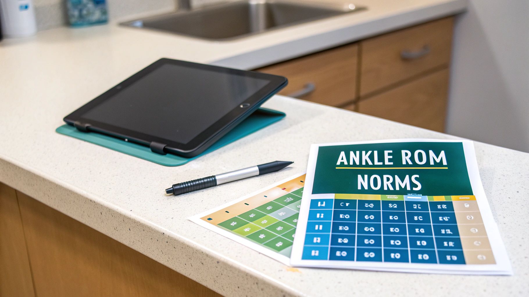
As a clinician, having a solid handle on ankle range of motion normal values is fundamental. These benchmarks provide a crucial baseline for everything from initial diagnosis to tracking rehabilitation progress.
For most adults, a typical range is approximately 20 degrees for dorsiflexion (pulling the foot up), 50 degrees for plantarflexion (pointing the foot down), 35 degrees for inversion (turning the sole inward), and 15 degrees for eversion (turning the sole outward) (1). These figures serve as a well-established starting point for any clinical assessment of the ankle.
Your Guide to Normal Ankle ROM Values
In physiotherapy and sports rehabilitation, range of motion (ROM) is a cornerstone metric. It quantifies the amount of movement available at a joint, offering objective insight into an individual's functional capacity and potential limitations.
For the ankle, these aren't just academic numbers. They directly translate to the ability to walk, run, squat, and navigate uneven ground safely. Establishing a patient's ROM against these norms is often a primary step after an injury, like an ankle sprain, which can account for a significant portion of all athletic injuries (2).
Why Standard Values Matter
Having a clear set of ankle range of motion normal values helps in several critical ways. First, it allows for the quick identification of deficits, whether it's hypomobility (stiffness) or hypermobility (excessive movement), which can lead to chronic instability. This is a common issue; a high percentage of individuals who experience an acute ankle sprain may develop long-term problems like chronic ankle instability (2).
Second, these norms guide the development of effective treatment plans. A patient with limited dorsiflexion requires a different therapeutic approach than someone with excessive, uncontrolled inversion.
Finally, tracking ROM over time is how progress is measured. This objective data is essential for justifying continued care, refining interventions, and making the crucial decision about when a patient is ready to return to their sport or daily life. To obtain this data, clinicians rely on various range of motion measurement tools to ensure accuracy.
Ankle injuries are among the most common in athletics. Comparing a patient's measurements to normative data helps a clinician create a clear pathway from injury back to peak performance.
To simplify, here is a summary of the generally accepted ankle range of motion normal values for healthy adults.
Summary of Normal Ankle ROM Values in Adults
This table presents the generally accepted average range of motion in degrees for the primary movements of the ankle complex, based on widely cited sources (1, 3).
| Movement | Average Range of Motion (Degrees) |
|---|---|
| Dorsiflexion | 20° |
| Plantarflexion | 50° |
| Inversion | 35° |
| Eversion | 15° |
It is important to remember that these are average values. They can vary based on factors like age, sex, and an individual's activity level, so sound clinical judgment is always paramount.
References:
- Soucie JM, Wang C, Forsyth A, Funk S, Denny M, Roach KE, et al. Range of Motion Measurements-Reference Values and a Database for American Adults and Children. The RARE Project. CDC; 2011.
- Vuurberg G, Hoorntje A, Wink LM, van der Doelen BFW, van den Bekerom MP, Dekker R, et al. Diagnosis, treatment and prevention of ankle sprains: update of an evidence-based clinical guideline. Br J Sports Med. 2018 Aug;52(15):956.
- American Academy of Orthopaedic Surgeons. The Clinical Measurement of Joint Motion. AAOS; 1965.
What Are We Actually Measuring? A Look at the Four Key Ankle Movements
Before discussing ankle range of motion normal values, it's essential to define what is being measured. The ankle is not a simple hinge; it is a complex of joints working in concert to allow movement in multiple planes. These motions are fundamental to everything we do on our feet.
The four primary movements are dorsiflexion, plantarflexion, inversion, and eversion. Each has a unique function in mobility and stability. While they often feel like a single fluid motion to a patient, clinicians understand they originate from distinct joints, a critical distinction for accurate assessment.
The Sagittal Plane: Up and Down
The most apparent ankle movements occur in the sagittal plane—the up-and-down motion of the foot. These actions are driven primarily by the talocrural joint, the articulation between the tibia, fibula, and talus.
-
Dorsiflexion: This is pulling the foot and toes up, closing the angle between the top of the foot and the shin. Sufficient dorsiflexion is essential for a normal walking gait, allowing the shin to move forward over the foot. It is also crucial for tasks like squatting or climbing stairs.
-
Plantarflexion: The opposite of dorsiflexion, this is the act of pointing the foot and toes down. Plantarflexion provides the propulsive force needed for walking, running, and jumping.
Together, these two movements provide the ankle's primary forward-backward mobility and are the foundation of human locomotion.
From a clinical standpoint, understanding why a movement is limited is paramount. For instance, is restricted dorsiflexion caused by a tight gastrocnemius, or is there an arthrokinematic restriction in the talocrural joint itself? The answer dictates the entire treatment plan.
The Frontal Plane: Side-to-Side Stabilizers
The ankle also moves in the frontal plane, allowing the foot to tilt inward and outward. These motions are our stabilizers, essential for maintaining balance and adapting to uneven ground. They primarily originate from the subtalar joint, located just below the talocrural joint.
-
Inversion: This is the motion of tilting the sole of the foot inward, toward the body’s midline. Inversion helps the foot conform to surfaces and plays a significant role in shock absorption upon foot strike.
-
Eversion: The counterpart to inversion, eversion involves tilting the sole of the foot outward, away from the midline. This movement is key for stabilizing the foot and creating a solid base of support.
These four movements provide a complete picture of the ankle's functional capacity. A solid grasp of their origin and function is the first step toward accurately measuring a patient's range of motion and interpreting the findings against normative data.
Mastering Standardized Ankle ROM Measurement
For clinicians, obtaining accurate and reproducible measurements is the bedrock of practice. For ankle range of motion, the goniometer remains a gold-standard tool, providing the objective data needed to guide treatment and demonstrate progress. Adherence to a standardized protocol is necessary to reduce variability between sessions or different clinicians.
This requires precise patient positioning, consistent goniometer placement, and the use of correct bony landmarks for alignment. Without this consistency, it is difficult to determine if a change in a patient's ankle range of motion normal values represents true clinical improvement or measurement error. Proper use of a goniometer is an essential skill; for a refresher, consider reviewing a detailed guide on what a goniometer is used for.
This visual guide breaks down the four primary movements of the ankle complex measured in clinical practice.
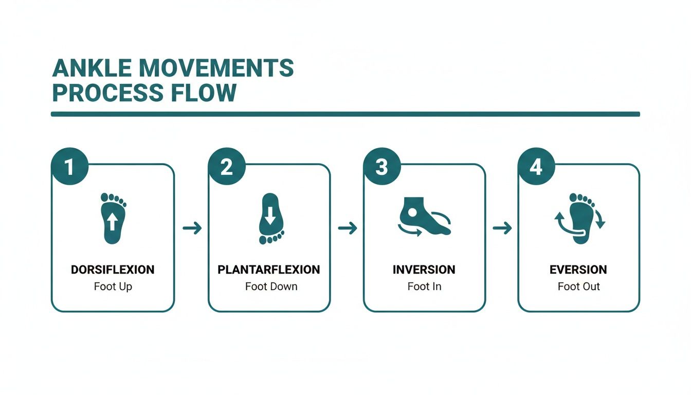
As shown, dorsiflexion and plantarflexion operate in one plane of motion, while inversion and eversion operate in another, highlighting the ankle's multi-axial nature.
Goniometric Measurement Protocol
To obtain a clean reading for any movement, a specific setup is needed to isolate the joint properly. Here is a step-by-step breakdown for measuring dorsiflexion and plantarflexion at the talocrural joint.
- Patient Position: The patient should be supine or seated with their knee bent to 90 degrees. This position reduces the influence of the gastrocnemius muscle. Crucially, the subtalar joint should be in a neutral position to prevent inversion or eversion from skewing the results.
- Goniometer Axis: Place the fulcrum of the goniometer directly over the lateral malleolus.
- Stationary Arm: Align the stationary arm with the head of the fibula.
- Moving Arm: Align the moving arm parallel to the shaft of the fifth metatarsal.
- Instruction: Ask the patient to actively pull their foot up (dorsiflexion) and then point it down (plantarflexion) as far as is comfortable. Record the degrees at the end of each movement.
The Weight-Bearing Lunge Test
While goniometry is excellent for assessing passive ROM, the weight-bearing lunge test (WBLT) provides crucial information on functional dorsiflexion. This test is highly relevant for athletes or anyone whose daily activities involve squatting or climbing, as it measures how far the tibia can travel over a planted foot.
The WBLT has demonstrated high reliability, with some studies reporting intraclass correlation coefficients (ICC) as high as 0.99 (4). Research indicates that while non-weight-bearing dorsiflexion averages around 16°, this can drop significantly in injured ankles. Functional, weight-bearing norms can be much higher, in a range that is vital for dynamic movements.
The key to mastering ankle ROM measurement isn't just knowing the steps—it's applying them with meticulous consistency. Every degree matters when making decisions about a patient's return to activity.
References:
- Bennell KL, Talbot RC, Wajswelner H, Techovanich W, Kelly DH, Hall AJ. Intra-rater and inter-rater reliability of a weight-bearing lunge measure of ankle dorsiflexion. Aust J Physiother. 1998;44(3):175-80.
Normative Data For Sagittal Plane Motion
When assessing mobility, solid, population-based data is the difference between guessing and making an informed clinical judgment. For the ankle, sagittal plane movements—dorsiflexion and plantarflexion—are fundamental to locomotion, so understanding typical ankle range of motion normal values provides a critical benchmark.
Large-scale studies provide a clear picture of how these values shift across a person's lifespan. It is well-documented that ankle mobility is not static; it naturally declines with age. This gradual reduction is a key factor to consider, especially when evaluating older adults or tracking a patient's joint health long-term.
Dorsiflexion and Plantarflexion Norms by Age and Sex
Research, including landmark data from the findings on ankle joint mobility from the CDC, offers detailed insights into these age-related changes (1). For example, dorsiflexion norms differ between a 25-year-old and a 45-year-old, with subtle differences between sexes.
This table breaks down some of these key differences, showing the expected decline with age.
Average Ankle Dorsiflexion & Plantarflexion by Age and Sex
| Age Group | Male Dorsiflexion (Avg) | Female Dorsiflexion (Avg) | Male Plantarflexion (Avg) | Female Plantarflexion (Avg) |
|---|---|---|---|---|
| 20-29 | 24.8° | 22.8° | 67.1° | 55.8° |
| 30-39 | 18.7° | 17.5° | 55.4° | 48.2° |
| 40-49 | 13.8° | 12.7° | 45.3° | 40.9° |
Data adapted from Soucie JM, et al. (2011) (1).
The decrease can be significant. For instance, average dorsiflexion for men in their 20s is 24.8°, falling to 13.8° by their 40s. Plantarflexion follows a similar pattern. Across the board, females tend to present with slightly less sagittal plane motion compared to their male counterparts in the same age group.
From a functional standpoint, these numbers represent capacity. A minimum of 10 degrees of dorsiflexion is widely considered necessary for a normal gait pattern. When an individual falls below this threshold, their body often compensates, which can lead to a cascade of issues in the knees, hips, and lower back.
This is why accurate assessment is critical. If you wish to refine your measurement technique, our guide on how to measure ankle dorsiflexion breaks down the clinical protocol.
Translating Data Into Clinical Practice
Knowing these normative values allows for the quick identification of meaningful deviations. Is a 45-year-old's limited dorsiflexion the result of a recent injury, or does it fall within the expected range for their age? Comparing their measurement to established norms for their specific demographic provides essential context.
Ultimately, this data-driven approach transforms a simple goniometric measurement into a powerful diagnostic and prognostic tool. It helps set realistic rehabilitation goals, educate patients on their progress relative to population benchmarks, and deliver more precise, effective care.
Normative Data For Frontal Plane Motion
While the sagittal plane handles forward momentum, frontal plane motion is where stability and adaptability occur. This involves inversion (rolling the sole inward) and eversion (rolling the sole outward), movements primarily driven by the subtalar joint. These motions allow the foot to conform to uneven ground, absorb shock, and maintain balance—critical for both daily activities and high-level athletic movements.
Having solid normative values for these side-to-side movements gives clinicians a clear benchmark for assessing mediolateral stability. This is especially crucial when managing ankle sprains, which almost always involve excessive inversion and can dramatically alter these ranges, often leading to chronic instability if not rehabilitated properly.
Gender Differences In Frontal Plane ROM
As with sagittal plane motion, factors like age and sex influence frontal plane mobility. Research consistently shows notable differences between males and females in this plane. This data is vital for building a more nuanced clinical picture.
A comprehensive 2011 study aimed to establish clear normative values and found significant gender-based variations. The data showed that, on average, women's ankles tend to have greater flexibility. Specifically, inversion averaged 25.49° in women versus 20.52° in men. Similarly, eversion was measured at 16.60° for women compared to 13.61° for men (5). You can explore the full research on ankle motion norms to see the complete breakdown.
These figures suggest that women typically have more available rotational movement. While this extra range might be advantageous in activities requiring flexibility, it could also be a consideration for injury risk if not supported by adequate muscular strength and neuromuscular control.
When assessing a patient, these gender-specific norms are incredibly useful. A female athlete presenting with 25° of inversion is within the average range for her sex. However, the same measurement in a male patient might suggest hypermobility or ligamentous laxity requiring further investigation.
Ultimately, these ankle range of motion normal values for inversion and eversion are an essential piece of the clinical puzzle. They help contextualize findings, set realistic rehabilitation goals, and make confident decisions about when an individual is ready to return to their activities safely.
References:
- Norkin CC, White DJ. Measurement of Joint Motion: A Guide to Goniometry. 4th ed. F.A. Davis Company; 2009.
Interpreting ROM Findings In A Clinical Context
Simply comparing a patient's measurements to a chart of ankle range of motion normal values is only the first step. The true clinical skill lies in interpretation—understanding what those numbers mean for that specific individual. Raw data must be placed within the full context of their age, activity level, and clinical presentation.
This process goes beyond merely spotting a deficit. It demands a nuanced understanding of concepts like hypomobility (less than normal movement) and hypermobility (excessive movement). Each points toward different underlying issues and requires a distinct therapeutic approach.
The Importance Of Bilateral Comparison
Before drawing conclusions, one of the most powerful tools is the patient's other ankle. Always measure the uninvolved side first. This simple step establishes the patient's personal baseline.
If their "good" ankle has 25 degrees of dorsiflexion, that becomes a much more relevant target than a generic population average of 20 degrees. Significant asymmetry between limbs is often a more telling sign than a minor deviation from a textbook value. A difference of 5-10 degrees between sides can be clinically significant, pointing to a unilateral issue such as a past injury, muscular imbalance, or joint capsule restriction.
You can learn more about how baseline data is used by reading our overview of what is normative data in a clinical setting.
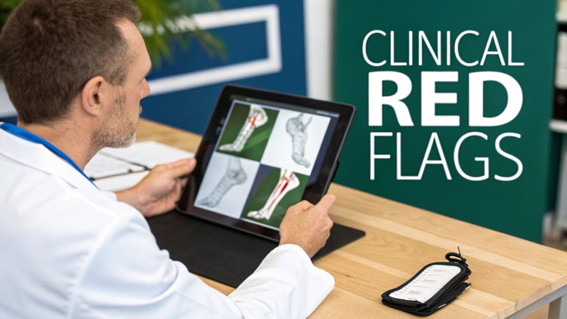
Red Flags In Ankle ROM Assessment
During assessment, certain findings should prompt further consideration. These red flags may indicate a more serious underlying pathology that needs further investigation or referral. Here, it is important to pay attention not just to the quantity of motion, but its quality.
Be alert for these key warning signs:
- Sudden, Painful Loss of Motion: A dramatic and painful drop in ROM without a clear traumatic event could suggest an inflammatory condition, a loose body in the joint, or another acute process.
- An "Empty" End-Feel: This occurs when the patient stops the movement due to intense pain before any real mechanical resistance is felt. It is a significant red flag for an active inflammatory process, a fracture, or other serious pathology.
- Spasm or Guarding: When muscles around the ankle fire uncontrollably to block movement, it is a protective response, often linked to an acute injury, significant joint instability, or severe inflammation.
A goniometer measures the quantity of motion, but a clinician's hands and eyes assess the quality. The end-feel—what is felt at the very end of the available range—provides crucial diagnostic clues about the cause of restriction, whether it's a hard bony block, a firm capsular feel, or soft tissue approximation.
When interpreting findings, it's crucial to consider the entire kinetic chain. For instance, incorporating exercises like stability ball exercises for a stronger core can be a vital part of a comprehensive rehabilitation plan, as a stable core contributes to better mechanics down to the ankle. This holistic view transforms raw data into a powerful tool for effective treatment.
Your Questions About Ankle ROM, Answered
Here are answers to some of the most common questions about ankle range of motion, connecting the measured values to their real-world implications for movement.
What's the Minimum Dorsiflexion I Need to Walk Normally?
For a smooth, efficient gait, at least 10 degrees of ankle dorsiflexion is generally required. This allows the shin (tibia) to move forward over the foot during the stance phase without the body needing to find a workaround.
When that 10 degrees is unavailable, the body often compensates, for example, with an early heel-off or by turning the feet outward ("out-toeing"). Over time, these compensatory patterns can add stress to the knees and hips.
Why Is My Ankle ROM Different When My Knee Is Bent vs. Straight?
This is a classic differential diagnostic technique used to determine which calf muscle may be restricting dorsiflexion. Measuring with the knee straight places the large gastrocnemius muscle under tension, as it crosses both the knee and ankle joints. If the range is limited in this position, the gastrocnemius is likely involved.
When the knee is bent to 90 degrees, the gastrocnemius is put on slack, effectively isolating other structures.
- If dorsiflexion improves significantly with the knee bent: This strongly indicates that tightness in the gastrocnemius is the primary limiting factor.
- If dorsiflexion remains limited with the knee bent: The restriction is more likely coming from the deeper soleus muscle (which does not cross the knee) or from stiffness within the ankle joint capsule itself.
Can I Actually Improve My Ankle Range of Motion?
Yes, in most cases, ankle ROM can be improved with a focused program of stretching and mobility exercises. If tight muscles are the cause, a targeted stretching routine for both the gastrocnemius and soleus is often the first step.
If the joint itself is the problem (a capsular restriction), a physical therapist can use manual therapy techniques, such as joint mobilizations, to help restore normal joint mechanics. A proper assessment from a healthcare professional is key to identifying the cause of the limitation and developing a safe, effective program.
A common mistake is performing only straight-knee calf stretches. While this targets the gastrocnemius, it is crucial to also perform stretches with the knee bent to properly address the soleus muscle and deeper joint structures.
What Else Affects Ankle ROM Besides Age and Gender?
While we have established ankle range of motion normal values, an individual's numbers are influenced by their activity level, injury history, and underlying medical conditions.
For example, a gymnast or dancer might be expected to have greater flexibility than textbook norms. Conversely, someone with a history of ankle sprains may show deficits, especially in dorsiflexion. Conditions like arthritis or certain neurological disorders can also significantly impact joint mobility. Therefore, ROM measurements should never be viewed in isolation but as part of a complete clinical picture.
At Meloq, we understand that objective, accurate data is the bedrock of good clinical practice. Our digital measurement tools are designed to help rehabilitation professionals replace subjective guesswork with precise, repeatable data. This empowers you to track progress with confidence and drive better outcomes for your patients. See how objective measurement can transform your practice at https://www.meloqdevices.com.

Featured Product
EasyAngle Digital Goniometer
Measure range of motion with clinical precision. CE certified, Bluetooth connected.
Learn More